Saturday, December 10, 2011

Pullorum Disease

Introduction


Disease caused by one of the two poultry-adapted strains of Salmonella bacteria, Salmonella Pullorum, this usually only causes mortality in birds up to 3 weeks of age. Occasionally it can cause losses in adult birds, usually brown-shell egg layers. It affects chickens most commonly, but also infects turkeys, game birds, guinea fowls, sparrows, parrots, ring doves, ostriches and peafowl. It still occurs worldwide in non-commercial poultry but is now rare in most commercial systems.

Morbidity is 10-80%; mortality is increased in stressed or immunocompromised flocks and may be up to 100%. The route of infection is oral or via the navel/yolk. Transmission may be transovarian or horizontal mainly in young birds and may sometimes be associated with cannibalism. The bacterium is fairly resistant to normal climate, surviving months but is susceptible to normal disinfectants.

Other Names


Bacillary white diarrhoea 

Organism


Salmonella Pullorum 

Signs


Inappetance.
Depression.
Ruffled feathers.
Closed eyes.
Loud chirping.
White diarrhoea.
Vent pasting.
Gasping.
Lameness.
Dejection.
Loss of appetite and thirst.
Closed eyes
Signs are generally mild compared to host-specific salmonellae, or absent.

Post-mortem lesions


Grey nodules in lungs, liver, gizzard wall and heart.
Intestinal or caecal inflammation.
Splenomegaly.
Caecal cores.
Urate crystals in ureters.

Diagnosis


Isolation and identification. In clinical cases direct plating on Brilliant Green and McConkey agar may be adequate. Enrichment media such as buffered peptone followed by selective broth or semi-solid media (e.g. Rappaport-Vassiliadis) followed by plating on two selective media will greatly increase sensitivity. However this has the potential to reveal the presence of salmonellae that are irrelevant to the clinical problem under investigation. Differentiate from Pullorum/Typhoid, other enterobacteria.
Differentiate from Typhoid, Paratyphoid, paracolon, other enterobacteria, chilling and omphalitis

Management Treatment


Eradication from breeder flocks. As with other salmonellae, recovered birds are resistant to the effects of infection but may remain carriers. Vaccines are not normally used as they interfere with serological testing and elimination of carriers.

Uninfected breeders, clean nests, fumigate eggs, all-in/all-out production, good feed, competitive exclusion, care in avoiding damage to natural flora, elimination of resident infections in hatcheries, mills, breeding and grow-out farms. Routine monitoring of breeding flocks, hatcheries and feed mills is required for effective control. Infection results in a strong immune response manifest by progressive reduction in excretion of the organism and reduced disease and excretion on subsequent challenge.Good management. Chemotherapy can prolong carrier status in some circumstances.

Physical Treatment


Amoxycillin, poteniated sulponamide, tetracylines, fluoroquinolones, Sulphonamides, neomycin, tetracyclines, fluoroquinolones.


Homeopathic Treatment


If birds huddle together & exhibit a White foamy diarrhoea give Calc carb+Cala phos

In Adult birds loss of appetite, weakness, greenish brown dirrhoea give Sulphur, Ipecac


Photo Gallery 


Pullorum disease is an acute systemic disease in chickens and turkey poults. The infection is trans¬mitted with eggs and is commonly characterized by a white diarrhoea and high death rate, whereas adult birds are asymptomatic earners. The morbidity and the mortality rates increase about the 7th - 10th day after hatching. The affected chickens appear somnolent, depressed and their growth is retarded. The feathers around the vent in many chickens is stained with diarrhoeic faeces or pasted with dry faeces.


Acute fowl typhoid. The outbreaks usually begin with a sharp decline in forage consumption and egg production. The fertilization and hatchability rates are considerably reduced. Diarrhoea appears. The death rate in acute fowl typhoid is high and varies between 10% and 90%. About 1/3 of chickens hatched from eggs from typhoid-infected flocks die. A characteristic lesion for acute fowl typhoid in adult birds is the enlarged and bronze greenish tint of liver.


Ureters are often filled with urates. For confirmation of the diagnosis, 5. pullorum should be isolated and typed. Pullorum disease must be differentiated from other salmonelloses, E. coli infections, Aspergillus that produces similar pulmonary lesions. Staphylococcus aureus, causing arthrites etc. Sometimes, the pulmonary nodes resemble the tumours in Marek's disease.


Chronic fowl typhoid. The lesions are primarily in the gonads. The ovaries are affected by inflammatory and degenerative changes.

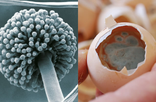


To keep the hatchery free of Aspergillus the following measures can be followed

Aspergillosis is a fungal infection of the respiratory tract in young birds, also commonly known as ‘brooder pneumonia’. In infected birds, the air sacs and lungs show white to yellow spots or lesions. Infected birds gasp for air and have ­accelerated breathing. Aspergillosis can also result in severe eye (and even brain) infection, which can appear as a yellow cheesy pellet beneath the eyelid. Increased mortality rates of 5 to 50 % may occur within 21 days after the placement of diseased birds.
Day-old chicks with signs of Aspergillosis are infected by the spores of Aspergillus species, among which Aspergillus fumigatus is the most common. The spores of the fungus Aspergillus are like small, dry seeds that can easily be spread by draught or the wind. The spores are found in low numbers throughout the whole environment. Aspergillus spores survive and grow in a wide range of conditions, but especially on organic matter, like egg yolk, cardboard boxes and wood. Growth in the spores is initiated by conditions of high humidity and temperature (37 - 45 ˚C). Cycles of high and low humidity optimise the growth of the fungus (mycelium) and the spread of its spores. The hatchery therefore provides optimum environmental conditions for Aspergillus to thrive.
Aspergillus spores can enter the hatchery either directly via the eggs, or via incoming air.
When the hatchery environment is contaminated by a high level of Aspergillus, the spores can easily enter the air ­handling unit and ventilation system. The climate, temperature and humidity in ventilation ducts is ideal for the propagation of Aspergillus, especially when organic matter (debris) has accumulated.
The main route into the hatchery for Aspergillus spores however, is usually via contaminated eggs. Aspergillus spores attached to the shell find their way into the albumen and yolk via hairlines or cracks in the shell. The yolk of the egg is an ideal substrate for the growth of Aspergillus and once the spores have entered the eggs, the way to the hatchery is open.
The nests also contain several sources of Aspergillus, including bedding, manure and feed. The nest eggs therefore become contaminated by contact with Aspergillus spores from these sources. Floor eggs also, of course, have a high risk of being heavily contaminated, when the spores enter the egg via hairline and other cracks.
Initially, infection by Aspergillus will be found during the routine analysis of unhatched eggs. Infected eggs show a visible mould on the membrane in the air cell (see picture). The risk of a serious bloom of Aspergillus is high when 0.5 % of the hatching eggs show clear infection with a visible growth of the fungus. Also, the frequency of embryos dying at about day 16 is higher then expected.
Advice A
Prevention is of course the first line of action. To prevent Aspergillus infection in the hatchery, the following measures are recommended:
Use Hatchery Recording Forms to ensure that the origin of eggs is traceable.
Do not incubate floor eggs.
Do not incubate cracked eggs or eggs with hairlines.
Avoid wooden walls, ceilings or surfaces in the hatchery, since Aspergillus thrives well on wooden surfaces,.
Analyse unhatched eggs on a regular basis - and if infected eggs are found, take measures to trace the sources of Aspergillus (see B).
Ensure your hatchery sanitation programme is fully maintained. If moulds are found, take immediate measures to clean the hatchery (see B).
Ensure that your hatchery sanitation programme includes the cleaning and disinfection of ventilation ducts.
Remember that hatchery equipment must be free of all organic matter before disinfection. It makes no sense to disinfect equipment, trays or boxes when debris remains stuck to the surfaces.

Advice B
If Aspergillus has been found in the hatchery the following is recommended:
Thoroughly clean and disinfect the hatchery - including ventilation ducts - with an effective fungicide. If necessary, ask your supplier for advice on the most effective solution.
Apply the fungicide at a regular interval because the spores of Aspergillus species are highly resistant to fungicides. Any surviving spores will develop into a mature spore-producing mould , thus the fungicide should be applied before this stage of development is reached.
Trace and eliminate the source of the Aspergillus spores, the main source of which is usually found at the breeder farm in the (wooden) nestboxes, litter, cardboard boxes and wooden walls or ceilings.

Aspergillosis in Poultry - Photo Gallery

 Photo Gallery:


1.Aspergillosis Is An Acute Or Chronic Respiratory Disease. In Rare Instances, Peritoneal, Visceral Or Systemic Lesions Could Be Observed. It Is Caused By Aspergillus Fumigatus. Dyspnea, Enhanced, Tense And Heavy Breathing Are Observed. Sometimes, Rales And Cyanosis Could Occur.
2.In The Acute Form, A Serous Fibrinous Pneumonia Is Observed. Within The Trachea And The Main Bronchi, Usually Near The Bifurcation, Obturation Masses Of Coagulated Fibrinous Exudate Are Detected.


3.4.In The Nodular Form, Multiple Grey Whitish Or Yellowish Dense Nodes In The Lungs Are Observed (379 And 380). Chickens, Turkeys, Waterfowl And Many Other Domestic Or Wild Fowl Are Susceptible.


5.6.7.Aspergillus Granulomas Of The Serous Coats: Pectoral (381), Abdominal Air Sacs (32) And Pleural (383), Are Sometimes Compressed. The Infection Could Occur Inside (Incu¬Batory Aspergillosis)Or Outside The Hatchery. Aspergillus Fumigatus Penetrates Through The Egg Shell Under Ideal Conditions For Development And Infects The Embryos. The Eggs Explode And Release A Significant Number Of Spores That Contaminate The Hatchery And The Environment. The Spores Are Spread Via The Venti¬Lation System That Results In Severe Outbreaks In Chickens Younger Than 3 Weeks. Outside The Hatchery, The Infection Occurs By Inhalation Of Numerous Spores From The Contaminated Forage, Litter Or Environment. The Overcrowding And The Increased Humidity Are Prerequisites For Outbreaks.


8.Rarely, As A Systemic Manifestation, Aspergillus Granulomas In The Brain Could Be Detected, When Spores Are Transported Via The Blood Circulation.

385
9.The Signs And Macroscopic Lesions Are Very Indicative For Aspergillosis. They Could Be Confirmed By Histology Too. In Acute Aspergillosis, Among The Inflammatory Necrotic Masses, The Spores Are Observed (Arrow A) As Well As The Grown Hyphae (Arrow B) Of The Mould.
388
12. Mould Is Developed After Inoculation Of Material On An Appropriate Nutrient Media.
13. Asperagellosis - Lung Nodules 


14. Aspergillosis, Brooder Pneumonia Lesions In A Chick.


                               15. Opened Left Abdominal Air-Sac Of Hen

Tuesday, December 6, 2011

Menacanthus stramineus


Menacanthus stramineus


Summary


Type: Chewing Lice
Zoonotic: No
Definitive Hosts: Birds
Inf Site: Skin (Epidermis)
Dx Tech: Direct Observation
Dx Stage: Adult with 6 Legs, Nymph with 6 Legs, Eggs("nits") on Hairs
Size: Macroscopic
Tran Mode: Direct Contact

Life Cycle


Lice undergo an incomplete metamorphosis which takes place entirely on the host within a period of 3-5 weeks. The eggs, commonly called "nits", are glued to the feathers of the host. The young lice develop through several nymphal stages, during which they look like small, pale adults. Nymphs and adults are transmitted from one animal to another primarily by host contact.

Taxonomy

Phylum Arthropoda
Class Insecta
Order Mallophaga
Suborder Amblycera

Diagnosis

Restlessness and anemia are two of the most outstanding clinical signs that should suggest pediculosis. Adults are small (up to 3.3 mm in length), with heads which are rounded anteriorly. The mouthparts are ventral and the claws are small. Careful examination of the skin and feathers of infested animals will reveal yellowish lice and their eggs which have distinctive filaments on the operculum and anterior half of the shell. Nits are laid in clusters.

Clinical Information

Chewing lice feed by nibbling on the feathers and epidermal debris, although some of them ingest blood by piercing the pulp of feathers or gnawing through the epidermis. Chewing lice annoy their hosts primarily by irritating the skin surface. A few lice are generally well tolerated, whereas large numbers may cause severe irritation, resulting in self-trauma. In general, lice are much more of a problem in young or debilitated animals than in older, well-nourished animals. A heavy louse infestation may itself be merely a symptom of some other underlying condition, such as malnutrition or chronic disease; debilitated animals often do not groom themselves and leave the lice undisturbed. 

Images

Cuclotogaster heterographus (Head Louse)


Cuclotogaster heterographus

SUMMARY


Type: Chewing Lice
Zoonotic: No
Definitive Hosts: Birds
Inf Site: Skin (Epidermis)
Dx Tech: Direct Observation
Dx Stage: Adult with 6 Legs, Nymph with 6 Legs, Eggs("nits") on Hairs
Size: Macroscopic
Tran Mode: Direct Contact


 Life Cycle


Lice undergo an incomplete metamorphosis which takes place entirely on the host within a period of 3-5 weeks. The eggs, commonly called "nits", are glued to the hairs or feathers of the host one at a time. Young lice leave the eggs by sucking air into their bodies and expelling it from the anus until they create a cushion of compressed air sufficient to pop open the lid of the egg. The young lice develop through several nymphal stages, during which they look like small, pale adults. Nymphs and adults are transmitted from one animal to another primarily by host contact. 


Taxonomy


Phylum Arthropoda
Class Insecta
Order Mallophaga
Suborder Ischnocera
Family Philopteridae


Diagnosis


 Restlessness and anemia are two of the most outstanding clinical signs that should suggest pediculosis. Careful examination of the skin and feathers of infested birds will reveal the lice and their eggs. Adults are 2 mm in length and greyish in color with chewing mouthparts.


Clinical Information


Chewing lice feed by nibbling on the feathers and epidermal debris, although some of them ingest blood by piercing the pulp of feathers or gnawing through the epidermis. Chewing lice annoy their hosts primarily by irritating the skin surface. A few lice are generally well tolerated, whereas large numbers may cause severe irritation, resulting in self-trauma. In general, lice are much more of a problem in young or debilitated animals than in older, well-nourished animals. A heavy louse infestation may itself be merely a symptom of some other underlying condition, such as malnutrition or chronic disease; debilitated animals often do not groom themselves and leave the lice undisturbed. Cuclotogaster heterographus is known as the "chicken head louse" because of its preference to settle on head and neck feathers of its host.


PHOTO GALLERY AND INFORMATION:


This illustration of female and male lice, Cuclotogaster heterog

This illustration of female and male lice, Cuclotogaster heterogThis illustration of female and male lice, Cuclotogaster heterographus shows the ventral aspect of this species (Nitzsh).
Cuclotogaster heterographus, or `chicken head louse', is a member of the group of chewing lice, which are small, flat, and wingless insects. This louse is a pest common to poultry, and feed on feathers, and scales of the skin.
CDC PHIL Image 5501

Figure 1 Cuclotogaster heterographus , ♀; Figure 2 Cuclotogaster heterographus , ♂

Sunday, December 4, 2011

Injured Chicken!

Injured Chicken!


When I got home today we found one of my beautiful chickens was laying injured amongst the leaves in the goat pen. She was limping on her good leg and then burrowing into the leaves. It was nice to see that one of her sisters refused to leave her side.


We brought her inside and examined her, it looked like she had a puncture wound and a possible broken leg. I put some neo-sporin on the open wound and gave her some chicken electrolytes with a bit of aspirin mixed in.


I didn't have any tongue depressors to use as a splint so I substituted some card stock as a replacement.


The card stock was covered with some vet wrap to hold it in place.


The chicken has been moved inside to Sara's kennel to see how she does. I read that injured chickens are attacked by their coop mates, I'm not sure if that is true but I don't want to take any chances.

I'm not sure if a chicken can survive a broken/injured leg, but I hope so.

Sara wants her kennel back and Patch, Mocha, and Chai Chai are very interested in our visitor, but they had better get used to it as the kennel may have to house kids and/or lambs this Spring.

Worms


Other Names


Internal Parasites


Organism or Mechanism


• Ascaridia galli (roundworm)
• Capillaria bursata, C. caudinflata, C. absignata,(roundworms)
• Syngamus trachea (nematode/gapeworm)
• Heterakis gallinarum (cecal worm)
• Cestodes Davainea proglottina
• Raillietina cesticullus (tapeworms)
• trematodes (flukes)

Preventative Measures


• Keep pastures clipped and limed.
• Graze pasture one year and cut the next.
• Use good hygiene practices.
• Rotate runs and houses.
• Keep age and species groups separate.
• Stack, heat, and sterilize poultry manure and do not spread it on poultry pastures.
• Avoid surface water, avoid damp places in houses.
• Leave runs empty for a least 2 months between batches.
• Allow grass to dry after dew or rain before allowing animals to graze.


Management Treatments


 • Remove infected litter and sanitize housing.
 • Plow and reseed infected fields.
 • If there is heavy contamination of earth-floored houses, remove the top 15-cm of soil.


Physical Treatments


• Give a 2-grain pill of santonin to each bird followed by 2 tsp. of raw linseed oil.
• For pinworms give a rectal injection of garlic, mustard, peppermint oil (1/2 tsp. oil/1 pint water), or eucalyptus (1/4 tsp./pint water) oil solution.
• Give Shakelee's Basic H surfactant administered as a solution in water.
•  Use Albomar 1ml for 1 kg body weight. This is available in India.

Nutritional Treatments


• Give a laxative diet: mash of pumpkin seeds and milk after a 12 hour fast, follow with a warm mash of bran, middlings, and milk.
• Give finely chopped onions and garlic.
• Feed finely grated raw carrot, garlic, onions, and parsley.
• Feed mustard; squash or pumpkin seeds; carrot and fennel seeds.
• Add DE and charcoal to feed all preceded by a fast and followed by a laxative period.

Herbal Treatments


• Give a course of garlic for ten days (1 clove/hen/day) and add to the diet bramble leaves, elder leaves, wormwood, wormseed, cotton-lavender, rue, hyssop,
• Give powdered cayenne or senna after a fast.
• If tapeworms are very bad give 1/2 tsp. grated male fern root mixed with bran, castor oil and molasses daily.
• Give garlic (pills, powders, fresh, tinctures),wormwood (Artemisia spp.), wild ginger, snakeroot, goosefoot, conifers (pine, spruce, firs), fennel seeds, or pyrethrum all preceded by a fast and followed by a laxative period.

• Verm-X is a Natural wormer with which you can remove the internal parasites.


Homeopathic Treatments


• If worms in the nostrils or ears causes circling give Cina.
• If affected birds lose weight, ruffled feathers, rapid breathing give--Aconite,
Santonite 3x, Tucrum merver.
• Head remedy--China, GNA.